Hydrocephalus: Understanding Diagnosis and Treatment
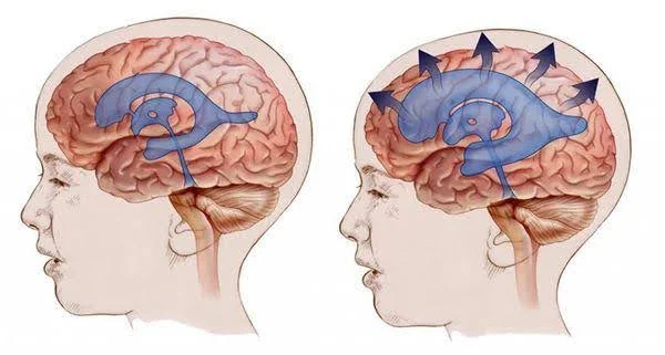
Hydrocephalus is a condition that occurs when there is an abnormal accumulation of cerebrospinal fluid (CSF) within the brain’s ventricles, cavities where this fluid is produced and circulates. CSF protects the brain, removes waste, and maintains a balance of nutrients. Under normal conditions, it flows continuously and is reabsorbed into the bloodstream; excess fluid creates pressure and can affect brain function.
Causes of hydrocephalus
There are several causes that can lead to hydrocephalus, including:
- Congenital: Conditions present at birth that affect CSF flow (e.g., spina bifida, brain malformations).
- Acquired: Developed after hemorrhages, infections, traumatic brain injuries, or tumors.
Types of hydrocephalus
- Communicating hydrocephalus: CSF flow is not blocked, but there is a problem with its reabsorption.
- Non-communicating hydrocephalus: Something obstructs CSF flow (tumor, cyst, congenital narrowing).
- Normal pressure hydrocephalus: More common in older adults; associated with walking difficulties, memory loss, and incontinence.
- Congenital hydrocephalus: Present at birth due to genetic malformations or problems in fetal development.
- Acquired hydrocephalus: Can occur at any stage of life due to injuries, infections, tumors, or brain bleeding.
Symptoms: How does hydrocephalus manifest?
- Babies: Increased head size (macrocephaly), bulging fontanel, irritability, vomiting, and feeding difficulties.
- Children and young adults: Persistent headache, nausea, visual problems, changes in balance, coordination, or behavior.
- Older adults: Unsteady gait or short steps, memory loss, confusion, and urinary incontinence.
If you experience any of these symptoms, consult a doctor.
Diagnosis: How do we confirm hydrocephalus?
- Clinical evaluation: Neurological examination and review of symptoms.
- Imaging studies:
- Computed tomography (CT): Detects CSF accumulation.
- Magnetic resonance imaging (MRI): Provides details of the ventricular system and possible causes of obstruction.
- Additional tests: Lumbar drainage or infusion tests to evaluate CSF management response.
Treatment: Available options
- Ventriculoperitoneal shunt: Tube that redirects excess CSF from the ventricles to the abdomen, where it is reabsorbed.
- Endoscopic third ventriculostomy (ETV): Minimally invasive procedure that creates a new pathway for CSF flow, avoiding shunt in cases of obstructive hydrocephalus.
- Medications: In some temporary cases, drugs that reduce CSF production.
- Treatment of primary cause: If hydrocephalus is secondary to infection, tumor, or bleeding, addressing the underlying problem is crucial.
Follow-up with a neurologist or neurosurgeon is essential to adjust the shunt system or management as the patient evolves.
Prognosis: What to expect?
With proper treatment, most patients show significant improvement. Some may require long-term monitoring or treatment adjustments, especially those with a shunt.
Conclusion
Living with hydrocephalus can present challenges, but with timely diagnosis, proper treatment, and continuous monitoring, many patients lead full and active lives. Learning about the condition, maintaining communication with healthcare professionals, and seeking support in patient groups contributes to a better quality of life.
If you have questions or need guidance, don’t hesitate to contact me for a specialized consultation.