Subdural Hematoma: What You Need to Know
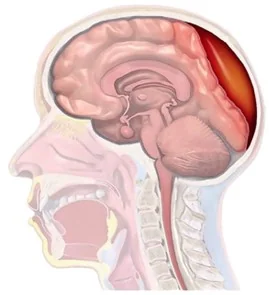
A subdural hematoma is an accumulation of blood in the subdural space, which is between the brain’s surface and the inner layer of the skull (dura mater). It can be a serious condition requiring immediate medical attention. This guide will help you understand what a subdural hematoma is, its causes, symptoms, and treatment options.
What is a subdural hematoma?
A subdural hematoma occurs when one or more blood vessels rupture in the subdural space, allowing blood to accumulate and put pressure on the brain.
Types of subdural hematoma
- Acute subdural hematoma: Appears shortly after severe head trauma and is usually very dangerous.
- Subacute subdural hematoma: Develops days or weeks after the injury.
- Chronic subdural hematoma: Forms slowly, often after a mild impact, especially in elderly people or those with blood clotting disorders; may go unnoticed initially.
Common causes
- Head trauma: Traffic accidents, falls, or high-impact sports.
- Neonates: “Shaken baby syndrome” after sudden movements.
- Advanced age: With aging, the brain shrinks and vessels become more fragile.
- Anticoagulants: Drugs like warfarin or aspirin increase bleeding risk.
- Chronic alcoholism: Weakens blood vessels.
- Spontaneous: Occur without a clear history.
Symptoms to watch for
- Persistent headache
- Decreased memory
- Nausea and vomiting
- Confusion or changes in mental state
- Excessive drowsiness
- Seizures
- Difficulty speaking, moving, or maintaining balance
- Loss of consciousness in severe cases
- Vision problems
If you or someone close to you experiences these symptoms after a head injury, seek immediate medical attention.
Diagnosis
Diagnosis is based on:
- Computed tomography (CT): Quick and effective method to identify bleeding.
- Magnetic resonance imaging (MRI): Useful for detecting older or smaller hematomas.
Treatment
Depends on the size and severity of the hematoma:
- Observation: Careful monitoring and repeat imaging studies in mild cases.
- Surgical drainage:
- Craniotomy: For large or complex hematomas; clot removal.
- Burr holes: Minimally invasive technique to drain chronic or small hematomas.
- Medications: To control intracranial pressure or prevent seizures.
Recovery and aftercare
- Regular monitoring: Medical check-ups to verify no new blood accumulation.
- Rehabilitation: Physical, occupational, or speech therapy as needed.
- Healthy lifestyle: Avoid alcohol, protect the head, and use anticoagulants only under medical supervision.
Prevention
- Use protective equipment in sports or risky activities.
- Avoid excessive alcohol consumption.
- Consult your doctor before taking anticoagulants.
- Maintain a safe home environment to reduce falls, especially for elderly people.
Conclusion
A subdural hematoma can be a serious condition, but with timely diagnosis and treatment, the chances of recovery increase significantly. If you have questions or concerns about this condition, don’t hesitate to consult a specialist.